This Technique Tuesday, our co-founder and equine veterinary surgeon, Jennifer Corley, is sharing her expertise on the injection of the Digital Flexor Tendon Sheath . Have questions or comments? Drop them in the comment form below, and Jennifer will be more than happy to help! Stay tuned for more exciting insights and techniques.


Indications
The superficial location of the digital flexor sheath means it is often penetrated by wounds and can become infected. Blocking the digital sheath is sometimes carried out in lameness examinations.
Equipment
- 20G 2.5-4cm (1- 1.5 inch) needle
- Scrub material
- Sterile gloves
- Clippers (optional)
10ml local anaesthetic if blocking
Anatomy
The digital flexor sheath encompasses the superficial digital flexor tendon (SDFT) and the deep digital flexor tendon (DDFT) from the distal third of the metacarpus/tarsus to just proximal to the navicular bursa. Distension of the sheath is easily differentiated from distension of the palmar/plantar fetlock joint pouch by location of the suspensory branch. Swellings in-front of the branch (dorsal) come from the fetlock. Swellings from behind the suspensory branch (palmar/plantar) are from the digital flexor sheath.
The palmar annular ligament, proximal digital annular ligament and distal digital annular ligament are effectively thickenings of the fibrous layer of the sheath wall which bind the flexor tendons to the palmar/plantar aspect of the limb. There are three levels to access the sheath each through gaps formed by these ligaments.
Proximal approach to the digital flexor sheath
A 20G 2.5-4cm (1-1.5 inch) needle is inserted at a level 3-4cm proximal to the lateral proximal sesamoid bone along the dorsal aspect of the DDFT. The needle is placed so that it is roughly between the DDFT and the lateral branch of the suspensory ligament. If there is no effusion it can be very difficult to obtain synovial fluid with this approach.
Mid-palmar/plantar approach (proximal collateral recess approach)
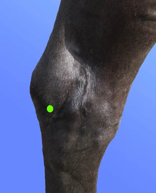
The proximal collateral recess is situated at the base of the sesamoid bone between the DDFT and the proximal insertion of the proximal annular ligament. A 20G 2-4cm (1-1.5 inch) needle is used. The site for needle placement is on the palmar/plantar aspect of the limb 1cm distal to the base of the lateral sesamoid bone and 1cm behind the neurovascular bundle. Deep palpation of the sesamoid bone allows a slight depression to be felt. The needle is directed at a 45 degree angle up towards this depression. This technique may be carried out with the leg lifted up or standing. Synovial fluid is usually obtained and has been shown to be most consistently obtained using the lateral recess with the leg raised.
Distal palmar/plantar approach
The largest pouch of the sheath lies between the branches of the SDFT over the distal pastern. At this between the branches of the SDFT and proximal digital annular ligament there is only skin overlying the sheath. A 20G 2-4cm (1-1.5 inch needle) is inserted at a level about 2cm proximal to the heel bulbs and 2-3cm off the midline of the palmar/plantar pastern. The needle is directed from the lateral towards the midline (horizontally across the pastern but aiming the needle downwards at a shallow angle so that the tip is aimed towards the middle of the pastern under the skin). Once placed the needle should be freely moveable and synovial fluid obtained. This technique is usually easier to perform with the leg picked up.
Preparation
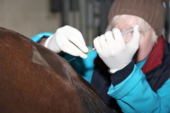
Prior to injection the site should be surgically prepared with an antiseptic soap such as polyhydroxy solution, povidone-iodine solution or chlorhexidine for a total contact time of seven or more minutes. Immediately before injection the skin preparation should be removed with 70% isopropyl alcohol. The horse can be sedated or restrained with a twitch.
Further resources
Equine Techniques App
https://apps.apple.com/us/app/equine-techniques/id451533346