This Technique Tuesday, our co-founder and equine veterinary surgeon, Jennifer Corley, is sharing her expertise on Epidural injection/catheterization. Have questions or comments? Drop them in the comment form below, and Jennifer will be more than happy to help! Stay tuned for more exciting insights and techniques.
-1.jpg?width=1200&length=1200&name=Epidural%20injecting%20(2)-1.jpg)
.jpg?width=1200&length=1200&name=epidural%20catheter%20(5).jpg)
Epidural injection/catheterization
Equipment
- Epidural needle 18 ga 5-9cm (2-3.5 inches) or epidural catheter kit.
- Clippers.
- Scrub material.
- Local anaesthetic.
- Surgical gloves.
- Drug to be injected.
- Elastoplast (for catheter).
- Sterile swabs.
- Sedation.
- Twitch.
Procedure
The horse needs to stand still and square. Stocks can be useful to protect from reflex kicks.
Anatomy for site for collection.
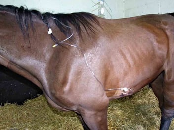
The site for epidural injection is between the coccygeal vertebrae 1 and 2 (C1-C2). The site is identified by raising and lowering the tail whilst feeling for the depression between the dorsal spinous process of the last immovable vertebrae (C1) and the first moveable vertebrae (C2). This location is usually about 5cm (2 inches) above the start of the tail hairs. Throughout the procedure the horse should stand squarely to help identify the midline.
The area is clipped and aseptically prepared. Local anaesthetic is infiltrated into the skin and subcutaneous tissue. If a catheter is to be placed local anaesthetic is required at two sites; at the catheter entry site and to the left or right of that site for suture to secure it.
Technique for injection
The successful angle of insertion of the needle depends on the anatomy of the horse and the relative positions of the skin puncture and the space between the vertebrae. Initially the needle is placed at ninety degrees and advanced until bone or ligament is encountered. If bone is encountered the needle is redirected at a lesser angle. Feeling penetration of the ligament may be very difficult.
Two methods can be used to determine if the needle is in the epidural space. The needle is advanced until the floor of the vertebral canal is encountered. As the needle passes past the nerve roots the tail should twitch. The needle must be withdrawn slightly to place the needle tip freely in the epidural space.
Alternatively a drop of sterile saline can be placed in the hub of the needle and the needle is advanced until the drop is sucked into the epidural space (this is called the ‘hanging drop’ technique).
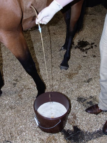
Technique for catheter placement
If a catheter is to be placed a small skin incision is made with a scalpel to allow the curved tip of the needle to slide through the skin. The needle is then advanced as before although it can be helpful to place the needle at a less steep angle to the skin to allow the catheter to more easily slide out of the needle. This requires a more caudal skin puncture. The absolute test for correct needle placement is that the catheter will advance. Some redirection and retraction of the needle might be needed to free the tip of the catheter as it passes out of the needle. Partially withdrawing the catheter stylet before advancing the catheter around the curve in the needle will facilitate stylet removal later. Care should be taken to direct the catheter craniad as indicated by the flat surface of the hub of the epidural needle. Once the catheter enters the epidural space it may slide forward as intended but it can also go caudal, circle or knot. For this reason it is prudent to only introduce enough catheter to prevent it from sliding or being pulled out (approx. 5cm). The needle is pulled off the catheter externally and the catheter stylet removed. To minimize the chance of unplanned removal and possible potential for descending infection the catheter is tunneled to 2-3cm under the skin from the site of entry. This is most easily done by making a second small skin incision to either side. The epidural needle should be passed from this to the catheter exit taking care not to cut the catheter. Insert the free end of the catheter retrograde through the needle and pull the needle and catheter through the subcutaneous tunnel to the new exit port. The excess catheter length should be trimmed and an injection port fitted to the free end. The catheter is secured with tape tabs, staples or sutures. The catheter site should be lightly bandaged for protection.
Injections should be given slowly particularly initially. Rapid injections of preservative free morphine and morphine with detomidine have precipitated muscle tremors , transient excitement and recumbency from which the horses immediately recover.
Epidural morphine can cause marked pruritus in a few horses, which can be largely be prevented by pretreatment with intramuscular acepromazine.