This Technique Tuesday, our co-founder and equine veterinary surgeon, Jennifer Corley, is sharing her expertise on the injection of the Calcaneal Bursa. Have questions or comments? Drop them in the comment form below, and Jennifer will be more than happy to help! Stay tuned for more exciting insights and techniques.
-1.png?width=1200&length=1200&name=MicrosoftTeams-image%20(1)-1.png)

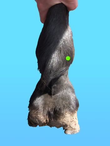
Indications and Anatomy
Wounds in the region of the plantar tarsus from the level of the mid-tibia to the distal inter-tarsal joint have the potential to penetrate and contaminate the bursae that lie over the point of the hock. There are three bursae that lie over the calcaneus in the area of the point of the hock;
- Subcutaneous calcaneal bursa (SCB)
- Intertendinous calaneal bursa (ICB)
- Gastrocnemius calcaneal bursa (GCB)
The nomenclature of the synovial bursae in this area can be confusing. The SCB and ICB have both been referred to as the bursa of the superficial digital flexor tendon (SDFT). Both the GCB and the ICB have been referred to as the subtendinous bursa. Many texts do not differentiate between the three bursa at all and refer to them only as the ‘calcaneal bursa’ which usually refers to the ICB. As the ICB and GCB do almost always communicate it is reasonable to think of them as one but clinically it is useful to understand the landmarks of both and appreciate that the openings are small and direct lavage of both structures may be required.
Anatomy
The anatomy of this area is variable and requires further study.
The SCB is located between the subcutaneous tissue and the superficial digital flexor tendon. Generally the centre is thought to be over the point of the tuber calcanei. However the distal extent may range from the proximal intertarsal joint to the most proximal extent of the tuber calcanei. The proximal extent can range from the proximal third of the tuber calcanei to the level of narrowing of the long digital extensor and lateral digital extensor muscle bellies.
The intertendinous calcaneal bursa lies between the superficial digital flexor tendon and the gastrocnemius tendon. While it usually extends 9.6cm proximal to the tuber calcanei and 7cm distal, it can have an extensive proximal limit reaching >15.6cm above the tuber calcanei.
The gastrocnemius calcaneal bursa lies beneath the gastrocnemius tendon dorsal to the insertion of the tendon on the calcaneus.
The SCB communicates with the ICB in around 39 % of horses. However given the extremely poor prognosis that sepsis of the ICB or GCB can have, it is always worth confirming lack of communication in the initial examination (this can be done by distending the ICB or GCB with saline).
The ICB and GCB almost always communicate, the exact anatomy of this communication has not been established but it is probably through small openings <10mm wide. While technically this would make the GCB a smaller compartment of the ICB clinically it is important to appreciate that the GCB may be a reservoir for sequestration of bacteria. Also that direct access to the GCB may be useful to treat animals where wounds prevent access to the ICB.
There is a lack of anatomical symmetry between bursa size, extent or communication between the left and right limbs in 78% of horses.
Preparation
Prior to injection the site should be surgically prepared with an antiseptic soap such as polyhydroxy solution, povidone-iodine solution or chlorhexidine for a total contact time of seven or more minutes. Immediately before injection the skin preparation should be removed with 70% isopropyl alcohol. The horse can be sedated or restrained with a twitch.
Equipment
20G 2.5cm (1 inch) needle
Scrub material
Sterile gloves
Clippers (optional)
10ml local anesthetic if blocking
Further Resources
Equine Techniques App
https://apps.apple.com/us/app/equine-techniques/id451533346
References
Burns J. Endoscopic instrumentation. In: Atlas of veterinary endosurgery Ed Slovis NM, Mosby, St Louis, 2003, pp 3-27
Holcombe SJ, Jackson C, Gerber V, et al: Stablin
g is associated with airway inflammation in young Arabian horses. Equine Vet J 33:244-249, 2001
Hardy J, Leveile R. Diseases of the guttural pouches. Vet Clin North Am Equine Pract 19:123-158 2003
Harold McKenzie Endoscopy of the guttural pouch in The Equine Hospital Manual, Blackwell 2008