This Technique Tuesday, our co-founder and equine veterinary surgeon, Jennifer Corley, is sharing her expertise on the Pleurocentesis. Have questions or comments? Drop them in the comment form below, and Jennifer will be more than happy to help! Stay tuned for more exciting insights and techniques.
.jpg?width=1200&length=1200&name=chest%20drain%20pleuropneumonia%20(1).jpg)
Indications
Shipping fever or other severe pneumonias can cause a build-up of fluid in the pleural space. This technique allows for the sterile collection of pleural fluid for cytological examination and culture.
Equipment
Catheter for centesis trocar 24Fr-28Fr x 41cm or teat cannula
Three way tap
Surgical gloves
Clippers with a fine (No. 40 blade)
Scrub material, chlorhexidine or similar as well as alcohol
No 15 scalpel blade
Local anaesthetic (2% mepivacaine) 5ml in a syringe with a 23-25 gauge needle
Ultrasound machine (optional)
One way valve or condom
Elastoplast
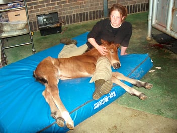
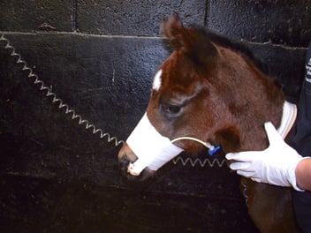
Twitch
Sedation
Anatomy
Selection for the site for pleurocentesis is ideally found by ultrasonography. Alternatively the procedure is performed in either the sixth or seventh intercostal space at a level approximately 10cm dorsal to the olecranon. Care must be taken to the lateral thoracic vein when performing the procedure. The intercostal blood vessels run down the caudal border of each rib and must be avoided. Potential complications of this procedure include pneumothorax and you should be prepared to deal with this (see next weeks blog). There may also be iatrogenic damage to the intercostal vessels, lung, heart or diaphragm.
Technique
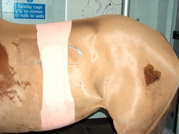
Care should be taken to avoid air aspiration after the catheter or cannula enters the pleural space. If using a trochar catheter have a hemostat or way to be able to close off the tube to hand for after stylet withdrawal. If using a teat cannula, a three way tap can be attached.
The insertion site is opened by making a stab incision in the skin. The skin incision is placed in the middle of the rib which is behind the intercostal space you have chosen.
The chosen site is clipped and aseptically prepared, followed by infiltration of the area with local anaesthetic. The chosen site is clipped and aseptically prepared, followed by infiltration of the area with local anaesthetic. The trochar catheter or is passed through the stab incision and directed forward around the cranial border of the rib caudal to the intercostal space being entered. The catheter is then aimed through the muscle at an almost perpendicular angle. Resistance will be encountered when going through the muscle and pleura which can be overcome with steady pressure. If additional force is required, use one hand to brace the catheter and minimise the risk of penetrating too deeply. Once it is felt the catheter has gone through into the peritoneal space (a small pop might be felt) the stylet should immediately be withdrawn a centimetre to avoid accidental damage to the lung or heart. Only the soft part of the catheter is then advanced.
A sample of fluid is taken using a Luer tip syringe, aliquots of the fluid are placed into EDTA and plain tubes for cytological examination and culture. Samples should be refrigerated until they are submitted to the diagnostic laboratory. If a large amount of fluid is present a trocar cannula may be sutured into place using a Chinese finger trap suture. The catheter should be closed between drainage episodes to prevent a pneumothorax. Commercial valves for chest drains are available alternatively a simple valve can be fashioned by attaching a condom securely over the end of the cannula. A small split in the end of the condom allows fluid to drain without air aspiration.