Technique Tuesday - IV catheters in foals
This Technique Tuesday, our co-founder and equine veterinary surgeon, Jennifer Corley, is sharing her expertise on IV catheters in foals. Have questions or comments? Drop them in the comment form below, and Kevin will be more than happy to help! Stay tuned for more exciting insights and techniques.
Indications
Sick foals often require intravenous fluids or antibiotics. Proper placement of an intravenous catheter is essential to deliver these. Catheters can be a source of infection. Proper aseptic technique during placement, careful wrapping, and daily site checking are crucial.
Suitable catheters
· 7Fr double-lumen (14-gauge/18 -auge) 20cm (8-inch) over-the-wire catheter.
· 16-gauge single lumen 20cm (8 inch) over-the-wire catheter.
· 16-gauge 13cm (5.25 inch) over-the-needle polyurethane catheter.
· 16-gauge 7.5cm (3 inch) over-the-needle polyurethane catheter.
Other equipment
Clippers with a fine (No. 40) blade
Drape or disposable incontinence pad (plastic-backed sheet)
Surgical masks
Surgical gloves
Scrub material, chlorhexidine and alcohol
2-0 non-absorbable monofilament suture on a straight needle
10ml heparinised saline (two syringes of heparinised saline if a double lumen catheter)
No 15-scalpel blade
Local anaesthetic (2% mepivacaine) 2ml drawn up in a syringe with 23-25G needle.
High-flow extension set
Injection cap
Adhesive bandage, 7.5cm Elastoplast
Zinc oxide tape
Bandage scissors
Anatomy
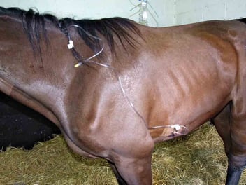
In foals, the jugular vein is usually used for catheterisation. The catheter should be placed around the border between the cranial and middle third of the jugular vein. The jugular vein has a bifurcation behind the angle of the jaw, which must be avoided. Placing the catheter too high can make it awkward to place the extension set. Catheters placed too low make it more difficult to identify correctly and increase the risk of putting the catheter into the carotid artery.
Technique
The hair should be removed from a rectangle 15cm long and 10cm wide over the jugular groove centred over the site selected for the cannula. A drape or disposable incontinence pad should be placed underneath the neck of the foal. The area is aseptically prepared. A 1ml subcuticular bleb of mepivacaine is placed at the site the catheter will be placed. A second bleb should be placed to allow the extension set to be sutured in place 3-4cm dorsal to the catheterisation site. Ideally, everyone holding the foal or assisting should put on surgical masks at this point. Much more excellent care is required for asepsis in foals than in adult horses. Foals are more vulnerable to sepsis and have fewer alternatives for sites for catheterisation if there is phlebitis. The clipped area is scrubbed again; some clinicians will even place sterile drapes in a four-point draping pattern. The procedure should always be performed using sterile gloves.
Placing an over-the-wire catheter
1. Prepare the Needle and Vein- Remove the J-wire from the kit and position it at the tip of the plastic nozzle.
- Hold the introducer needle between the thumb and forefinger of your right hand.
- Use your left first finger's knuckle to gently elevate the vein in the clean area.
2. Inserting the Needle
- Keep the needle parallel to the jugular vein, angled at 35 degrees to the skin.
- Advance the needle until blood appears.
- Adjust the angle slightly (5-10 degrees) and insert it further (1-2cm).
- Hold the needle steady with the left hand while maintaining vein elevation to prevent air intake.
3. Using the J-Wire
- Without altering the needle's position, insert the J-wire into the needle using your right hand.
- Advance the wire using your thumb. It should move smoothly in a correctly positioned needle.
- If the wire stops after 6-7cm, the needle might be out of the vein. Remove the wire and replace the needle.
4. Remove the needle and use the skin dilator
- Advance the wire until 5-10cm is left outside the needle. Securely hold the wire's end to prevent it from getting lost in the vein or touching unprepared skin.
- Remove the needle while holding the wire with the right hand.
- Insert the skin dilator over the wire into the vein. Rotate it slightly and push, overcoming resistance in the tissue.
- Move the dilator to the hub and withdraw it carefully.
5. Insert the catheter along the wire and secure it
- Hold the wire at a near 90-degree angle to the foal’s neck.
- Carefully slide the catheter onto the wire. Ensure the wire emerges from the catheter port by adjusting if necessary.
- Seat the catheter fully against the skin and withdraw the wire about 5cm.
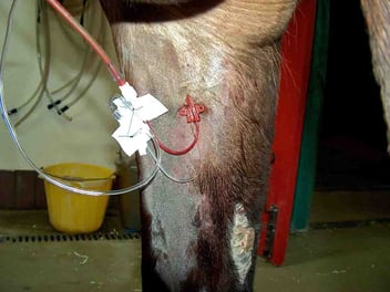
- Securely suture the catheter in place.
6. Post-Catheterization Care
- Withdraw and discard the wire.
- Optionally, take a blood culture sample, flush the catheter, and suture the extension set.
- Use zinc oxide tape to secure the extension set and bandage the catheter assembly gently to avoid vein obstruction.
Placing an over-the-needle catheter
Preparation, including placement of local anaesthetic, is as previously described. The catheter should be freed up with sterile gloves by sliding 2-3mm down the needle and back up again.
With some catheters or foals, a small skin incision may be helpful. The catheter is placed with the hub towards the head of the horse.
The catheter is advanced into the vein at 45 degrees to the foal’s neck. Once a flash of blood is seen, the catheter is flattened towards the neck of the foal at 5-10 degrees. The catheter is advanced an additional 0.5cm-1.5cm. If blood is still coming back, the catheter is slid into position.
An extension set or injection cap is placed immediately, and the catheter is sutured into position. The catheter should be wrapped as an over-the-wire catheter.
Catheter care
Twice daily checks for heat and swelling are essential. The vein should be monitored for any heat, swelling or hardness. If any part of the catheter apart from the hub is exposed, it should be re-sutured to prevent this. This type of movement can result in bacteria from the skin being tracked into the vein. If there is resistance to flow through the catheter, it should be closely inspected. Extension sets can move and kink. The catheter may also kink just beneath the hub. If this occurs, it must be replaced. Catheters that are removed for suspected infection should be cultured.