This Technique Tuesday, our co-founder and equine veterinary surgeon, Jennifer Corley, is sharing her expertise on the Cephalic Catheter. Have questions or comments? Drop them in the comment form below, and Jennifer will be more than happy to help! Stay tuned for more exciting insights and techniques.


Indications
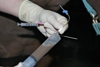
If there is thrombosis in one jugular vein, it is not recommended to use the other. The cephalic vein offers an alternative site for IV treatments where necessary. Placing a catheter in the foreleg is potentially dangerous to the veterinarian. Not all horses will tolerate this procedure. The cephalic is a small vein and the maximum rate of fluid that can be administered in an adult is 5L/hr. Over the wire catheters are easier to place, they are less stiff and less likely to kink.
Suitable catheters
· 14G single lumen 20cm (8 inch) over the wire catheter
· 16G single lumen 20cm (8 inch) over the wire catheter
· 14G 13cm (5.25 inch) over the needle polyurethane catheter
· 16G 13cm (5.25 inch) over the needle polyurethane catheter
Other equipment
Clippers
Scrub material
2-0 nonabsorbable monofilament suture on a straight needle
30ml heparinized saline
No 15 scalpel blade
Local anaesthetic (2% mepivacaine)
2ml syringe with 25G 1.6cm (5/8 inch) needle
Extension set
Injection cap
Adhesive bandage
Sterile gloves
Zinc oxide tape
Bandage scissors
Sedation (optional)
Twitch (optional)
Ultrasound (optional)
Anatomy
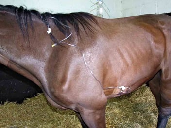
The cephalic vein runs along the medial aspect of the foreleg. The catheter is placed halfway between the carpus and the elbow.
Technique
The area for placement of the catheter is clipped and asceptically prepared. The catheter is placed pointing cranially (hub caudal, tip cranial). Local anaesthetic is used, place this both where the catheter will be placed but also at the site the extension set will be attached. The catheter is placed with the hub towards the carpus and the tip towards the elbow. The vein is raised and the needle place at a flat angle. The vein is very superficial. The needle does not need to be advanced to the hub. The wire is fed into the needle and then the over the wire catheter fed in using the standard technique. Once placed and sutured the catheter hub is covered with a dry swab and the whole catheter wrapped with an adhesive bandage.