This Technique Tuesday, our co-founder and equine veterinary surgeon, Jennifer Corley, is sharing her expertise on Tracheal Aspiration. Have questions or comments? Drop them in the comment form below, and Jennifer will be more than happy to help! Stay tuned for more exciting insights and techniques.
.jpg?width=1200&length=1200&name=Tracheal%20Aspirate%20for%20Blog%20(1).jpg)
.jpg?width=1200&length=1200&name=Tracheal%20Aspirate%20for%20Socials%20(1).jpg)
Endoscopic technique
Endoscope (cleaned and disinfected)
Guarded aspiration catheter
Endoscopic light source
20ml sterile saline
Percutaneous (trans-tracheal) technique
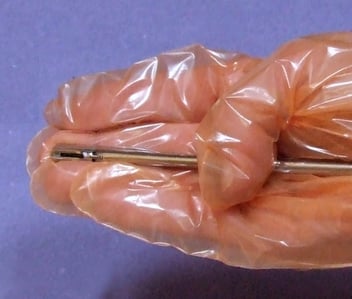
Trans-tracheal kit (commercial)
12 ga IV catheter (7.5cm/ 3inch) or 12 ga needle (7.5cm /3 inch) or nesting trocar
Polypropylene catheter (5-6F dog urinary catheter)
Clippers
Scrub material
2 x 3ml local anaesthetic with 25 ga needle
40 ml sterile saline
Surgical gloves
Required for both techniques
Sterile plain pot or tube
Sample pot with EDTA
Sedation (optional)
Twitch (optional)
The endoscopic technique described is less invasive but more likely to lead to sample contamination. The trans-tracheal technique is associated with a number of complications) subcutaneous emphysema, cellulitis tracheal damage pneumomediastinum) but is more appropriate especially if a sample for bacteriological culture is required. If both endoscopy and a sample for bacteriological culture are required bacteriological samples should be taken first.
Transendoscopic technique
The endoscope must be thoroughly cleaned and flushed prior to taking the sample to avoid contamination. Even with thorough cleaning many scopes retain a bacteriological population which can confuse diagnosis. The scope is advanced through the larynx to the mid cervical trachea or until a small pool of fluid in the cranial trachea is seen. The transendoscopic technique uses a double or triple stage guarded system of tubing. This is introduced into the tracheal lumen using the endoscopic biopsy channel. Fluid is then aspirated using a sterile syringe. If no fluid is present 20 ml sterile fluid is injected into the trachea and then aspirated. The sample should be placed into sterile plain and EDTA tubes for immediate transport to the laboratory. The sample should ideally be kept refrigerated in transit to avoid cellular degeneration.
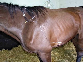
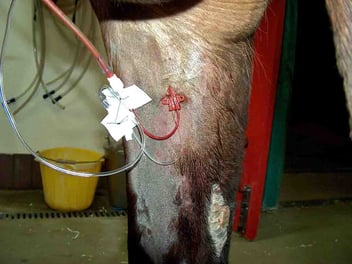
Transtracheal technique
The site for percutaneous aspiration is in the mid-cervical region on the midline. The skin overlying the trachea is anaesthetized with 2-3ml of local anaesthetic. The procedure is performed using a commercial kit. If a kit is not available an IV catheter and dog urinary catheter may be used. Check in advance that the IV catheter is of sufficient diameter for the dog urinary catheter to pass through. A 6F dog urinary catheter usually fits a 12 gauge catheter. The trachea is palpated and the catheter is then pushed between two tracheal rings about 2cm into the lumen of the trachea. The urinary catheter is then fed down into the trachea to the level of the thoracic inlet. 3ml local anaesthetic is injected first to reduce coughing which can contaminate the sample. 30-40ml of sterile saline is then injected in and immediately aspirated. On completion of aspiration it is very important to withdraw the urinary catheter prior to removing the catheter. Removing the catheter simultaneously risks seeding infection into the overlying skin.