This Technique Tuesday, our co-founder and equine veterinary surgeon, Jennifer Corley, is sharing her expertise on the Evacuation of Pneumothorax. Have questions or comments? Drop them in the comment form below, and Jennifer will be more than happy to help! Stay tuned for more exciting insights and techniques.


Indications
Pneumothorax (air in the pleural cavity) can occur after trauma, secondary to severe pneumonia or be iatrogenic. Pneumothorax is a potential complication of pleurocentesis. Unilateral pneumothorax is quite well tolerated in the horse but if the thin fenestrated caudal mediastinum breaks down then bilateral pneumothorax will ensue which is potentially fatal. Symptoms of pneumothorax include respiratory distress, tachypnea, flaring nostrils with hypoxaemia and cyanosis in severe cases.
Required Equipment
Catheter for centesis; trocar 24Fr-28Fr x 41cm, or teat cannula, or 7.5cm 18G spinal needle.
Method to confirm pneumothorax
X-ray machine capable of 80-90 kV and 40mA-s with grid
Ultrasound machine with a 3-7.5 MHz sector, linear array or linear probe.
Surgical gloves
Clippers with a fine (No. 40 blade)
Scrub material, chlorhexidine or similar as well as alcohol
No 15 scalpel blade
Local anaesthetic (2% mepivacaine) 5ml in a syringe with a 23-25 gauge needle
One way valve or three way tap
Valved test tube adapter
Elastoplast
Twitch
Sedation
Anatomy
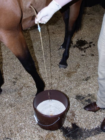
The finding of pneumothorax is ideally found by radiography or thoracic ultrasonography. The procedure is performed in a similar way to pleurocentesis (see last weeks blog post). However the site should be higher up, typically at the level of the point of the shoulder. Small amounts of air may be aspirated using a 7.5cm 18 gauge spinal needle or teat cannula. A three-way stopcock or centesis valve should be placed on the needle or cannula prior to insertion in order to prevent more air being sucked into the pleural cavity. Once in position the air can be removed using a 60ml Luer lock syringe. Using a needle or teat cannula does not allow for ongoing evacuation of air. If an indwelling solution is likely to be needed then an indwelling thoracic trocar catheter is usually preferred. The catheter should be sutured in place and fitted with a one way valve. Horses with a complete mediastinum may need bilateral catheters to be placed.
Watch the video: